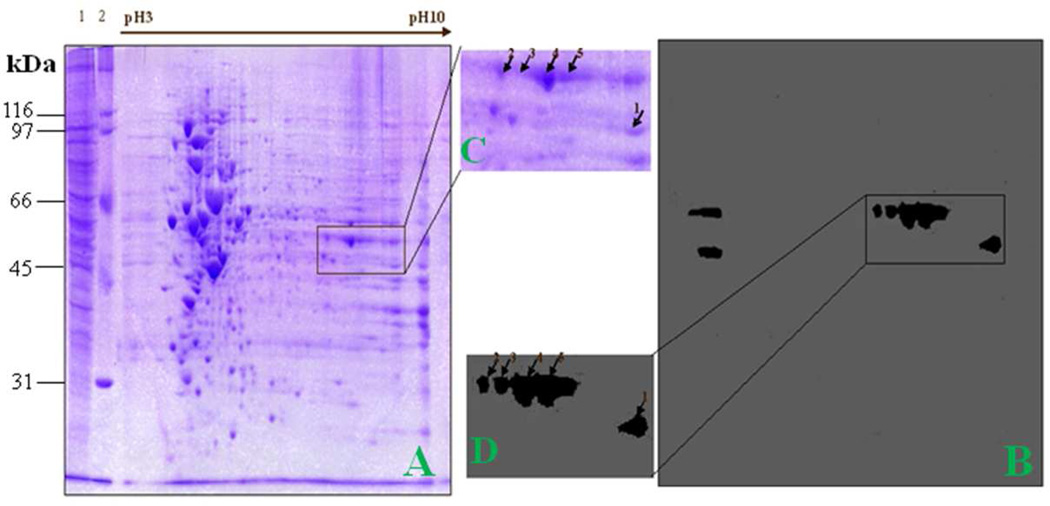
Figure 4. Identification of the 47-kDa liver fibrosis-associated protein by immunoproteomics.
(A) The 2DE protein profile of HepG2 cells. Lane 1, HepG2 cell lysate; lane 2, protein molecular marker. (B) Western blotting analysis of 2DE gel was probed with a high-titer liver fibrosis serum which contains antibodies to the 47-kDa protein. (C) and (D) The corresponding area (rectangle) of A and B was enlarged. Positive spots were picked up by using PDQUEST software.

