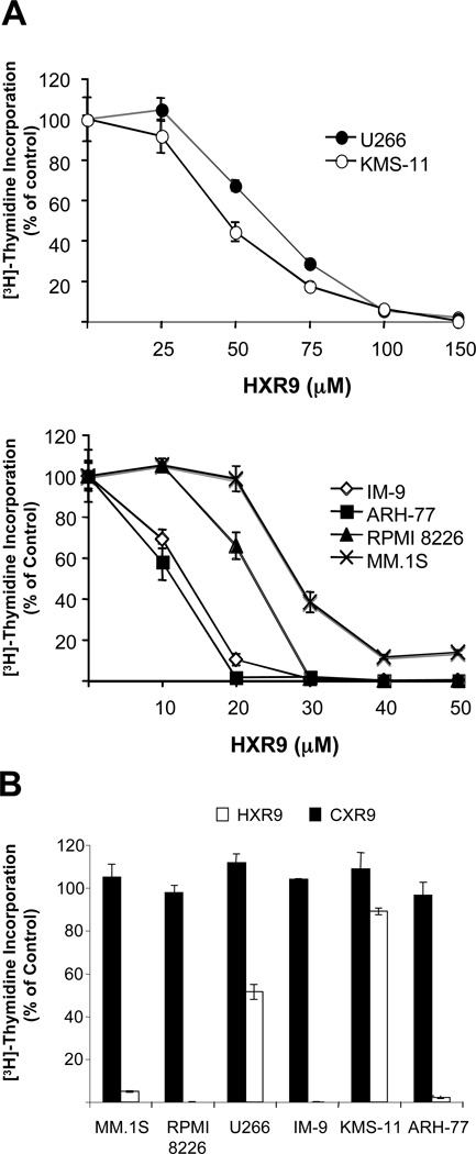
Figure 2. Anti-proliferative effects of HXR9 in malignant B cells.
A) Cells were treated in triplicate with various concentrations of HXR9 for 48 hours. The [3H]-thymidine incorporation assay was used to monitor cell proliferation. Least sensitive cell-lines, U266 and KMS-11 (top panel); sensitive cell lines, IM-9, ARH-77, RPMI 8226, and MM.1S (bottom panel). B) Specificity of HXR9 was confirmed by testing all cell lines with the control peptide CXR9. Cells were treated with 50 µM CXR9 or HXR9 for 48 hours. Data is expressed as a percent of the [3H]-thymidine incorporation of control cells. Standard deviation is indicated for each treatment. Data is representative of two independent experiments.

