Figure 1:
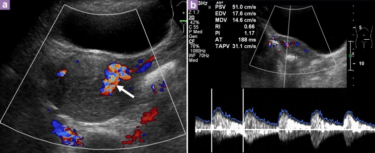
Transabdominal ultrasound of the pelvis shows bulky uterus with increased vascularity and multidirectional flow. Prominent vessel is seen on the left lateral wall of the uterus (arrow), likely to arise from the left uterine artery. b) Spectral Doppler showed a peak systolic velocity (PSV) of 51 cm/s and resistive index (RI) of 0.66.

