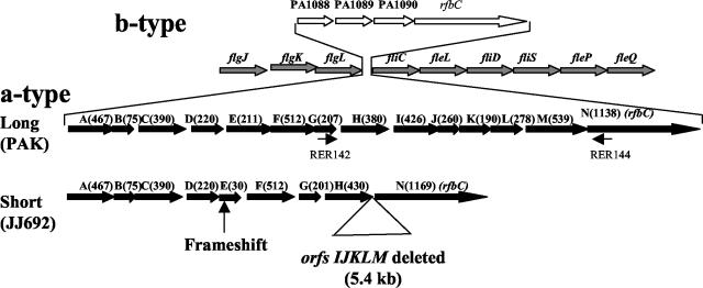
FIG. 3.
Schematic diagram showing the structure of GIs of a- and b-type P. aeruginosa strains. The diagram shows the region of the P. aeruginosa chromosome where the GIs are located. The location of the GI insertion is shown to be in the middle of flagellar genes (shown by arrows with gray filling) flgL on the 5′ end and fliC at the 3′ end. Two a-type GIs, long (PAK) with all 14 ORFs and short (JJ692) with a 5.4-kb sequence containing deletions of orfI, -J, -K, -L, and -M, are shown by black filled arrows. The PAO1 GI with 3 ORFs of unknown function and an ORF which had homology to rfbC gene (orfN) is shown by arrows with no fill for comparison. The locations of the primers RER142 and RER144 used for PCR analysis of different P. aeruginosa strains are shown by small arrows.

