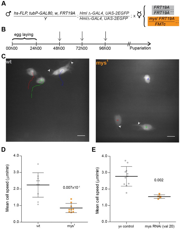
Fig. 1. Myospheroid is required for proper hemocyte migration.

(A) Outline of the MARCM protocol in hemocytes. Cross between DEMON males and FRT19a control or mys1 mutant virgin female flies for MARCM analysis. (B) Crosses are placed at 25°C for 24 hours. The progeny is submitted to three 1 hour heat-shocks (indicated by arrows) at 37°C before selection of 3rd instar females containing GFP-expressing hemocytes. (C) Movement of wild-type and mys1 GFP-expressing hemocytes in 3 to 4 hour APF flies, tracked for 12 minutes (1 min time-lapse interval). Arrowheads indicate filopodium-like protrusions produced by both wild-type and mys1 mutant hemocytes. Scale bars: 10 µm. (D) Graph showing individual mean cell speed for wild-type and mys1 clones (2 to 4 hours APF). (E) Graph showing average mean cell speeds for yv control and mys (valium 20) RNAi-expressing flies (between 3 to 4 hours APF). Mann–Whitney tests for non-Gaussian populations were used. Black lines indicate the samples' means; Error bars = standard deviation; P-values shown above tested groups.
