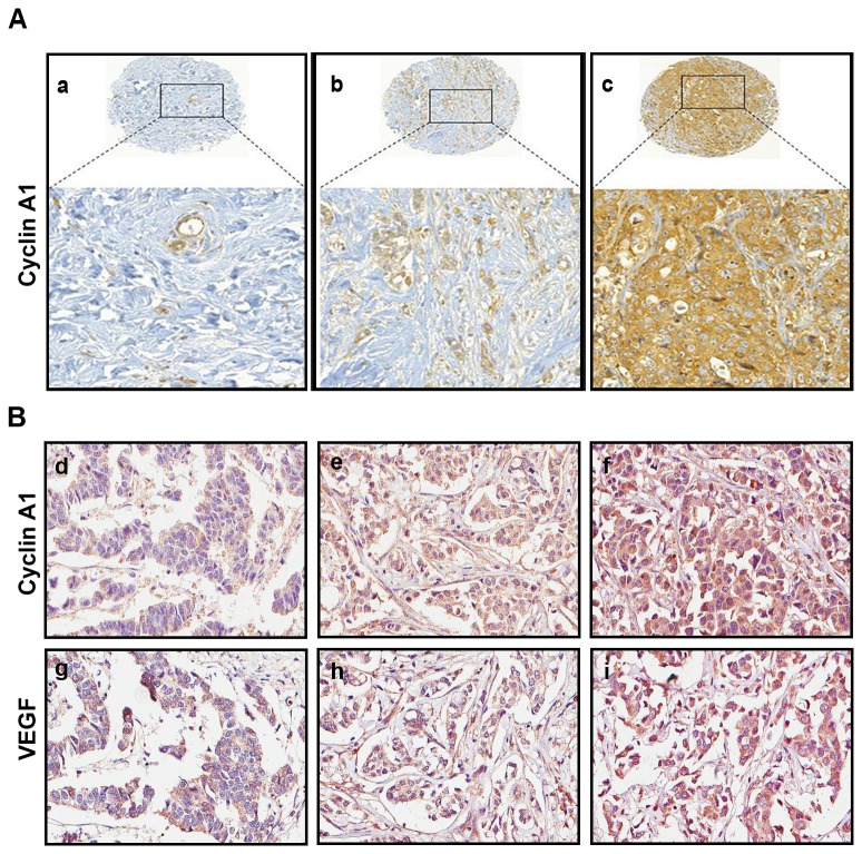
Figure 1. Evaluation of the expression of cyclin A1 and vascular endothelial growth factor (VEGF) in breast cancer specimens.
(A) Primary breast cancer specimens from TMA1 were immunostained with antibodies against cyclin A1. Representative microphotographs of TMA cores (upper panels) and the enlarged area (lower panels) showing cyclin A1 staining intensities from weak (a), moderate (b) to high (c) levels are presented. (B) Representative microphotographs show the staining intensity of cyclin A1, weak (d), moderate (e) to high (f) and VEGF, weak (g), moderate (h) to high (i) in primary breast cancer specimens from TMA2.

