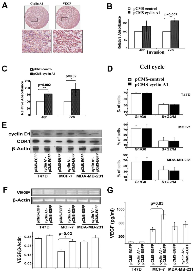
Figure 3. Effect of cyclin A1 expression on tumor invasion is associated with its effect on VEGF expression in MCF-7 cells.
(A) Evaluation of cyclin A1 and VEGF expression in metastatic lesions from lymph nodes from patients with breast cancer metastasis using immunohistochemical analysis. Representative pictures show the cancer cells are strongly positive to cyclin A1 and VEGF expression. Upper panels represent cores at 20x magnificantion and lower panels represent the higher magnification (40x) of the selected areas. (B) MCF-7 cells that were transfected with cyclin A1pCMS-EGFP or pCMS-EGFP vectors were applied on the Matrigel-coated invasion chamber and were assessed after 48 or 72 hours. Data in graphs are the mean ± SD represents two independent experiments, each performed in duplicates. P value is indicated. (C) MDA-MB-231 cells transfected with cyclin A1pCMS-EGFP or pCMS-EGFP were applied on the Matrigel-coated invasion chamber and were analyzed after 48 or 72 hours. Data in graphs are the mean of two independent experiments, each performed in duplicate, p=0.002 for 48 h and p=0.02 for 72 h. (D) Cell cycle distribution of the cells that were transfected with cyclin A1pCMS-EGFP or pCMS-EGFP. Data in graphs are the mean ± SD represents three independent experiments from flow cytometry analysis. The percentage of cells at onset of each cell cycle phase is indicated. (E) Western blot analysis shows the levels of cyclin D1 and CDK1 protein expression in the cells that were transfected with cyclin A1pCMS-EGFP or pCMS-EGFP. (F) Representative picture shows the VEGF mRNA expression in the cells transfected with cyclin A1pCMS-EGFP or pCMS-EGFP (upper panel). Quantification of VEGF mRNA level in the samples is indicated. P value is shown (lower panel). (G) ELISA assay of VEGF secretion in the cells transfected with cyclin A1pCMS-EGFP or pCMS-EGFP. Mean ± SD represents three independent experiments (lower panel). Breast cancer cell lines used for these studies are T47D, MCF-7 and MDA-MB231 as indicated.

