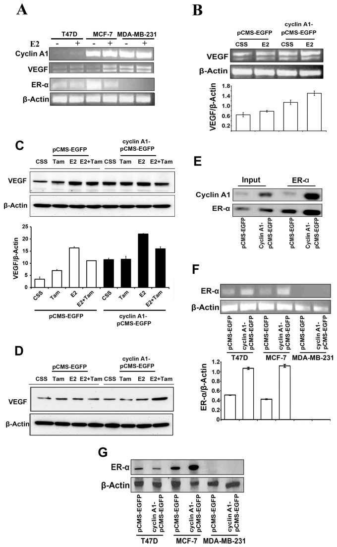
Figure 4. Evaluation of the association between cyclin A1 and ER-α estrogen signaling and the regulation of VEGF expression.
(A) -Evaluation of the effect of estrogen on mRNA expression of cyclin A1, VEGF and ER-α, in T47D, MCF-7 and MDA-MB231 cells using semi-quantitative RT-PCR analysis. The three cell lines mentioned above were cultured in charcoal stripped medium (CSS) or in CSS medium containing E2 at 5 µM for additional 48 hours as indicated. (B) The effect of E2 treatment or cyclin A1 overexpression alone or in combination on VEGF mRNA level was determined in MCF-7 cells. Cells that were transfected with cyclin A1pCMS-EGFP or pCMS-EGFP vectors in the absence or presence of E2 are indicated. (C) Evaluation of the effects of Tamoxifen (Tam), E2, and E2 in combination with Tamoxifen (E2+Tam) on VEGF protein expression in MCF-7 cells. MCF-7 cells were transfected with cyclin A1 vector or control vector as indicated. Data in graphs below are the mean ± SD represents two independent experiments. (D) Immunoblot analysis data obtained in MDA-MB-231 which were treated using the same conditions as mentioned in (C). (E) Immunoprecipitation analysis (IP) shows physical interaction between cyclin A1 and ER-α in MCF-7 cells that were transfected with cyclin A1pCMS-EGFP or pCMS-EGFP vectors. ER-α antibody was used in IP to pull down the immunocomplexes and subsequent Westernblot was performed using cyclin A1 or ER-α antibodies to detect the immunocomplexes as indicated. The input was used as controls as indicated. (F) Evaluation of ER-α mRNA expression in the cells that were transfected with A1pCMS-EGFP or pCMS-EGFP vectors. The representative picture is shown in the upper panel. Quantification of ER-α mRNA level is shown in the lower panel and mean ± SD represents three independent experiments. (G) Western blot analysis shows the expression level of ER-α protein in the cells that were transfected with A1pCMS-EGFP or pCMS-EGFP vectors. Breast cancer cell lines used for these studies are T47D, MCF-7 and MDA-MB231 as indicated.

