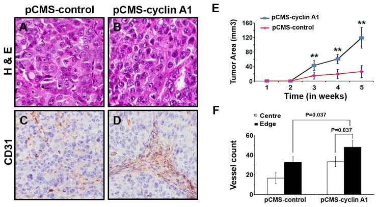
Figure 5. The effect of cyclin A1 on growth and vascularization of tumor xenografts in mice.
MCF-7 cells transfected with cyclin A1pCMS–EGFP or pCMS-EGFP vectors were subcutaneous implanted into female nude mice with E2 supplementation. (A, B) Representative microphotographs of xenograft tumor sections stained with Haematoxylin and Eosin are shown. The control tumor “pCMS-control” and cyclin A1 expressing tumor “pCMS-cyclin A1” are indicated. (C, D) Representative pictures show the xenograft tumors stained with antibody against human CD31, the CD31 positive vessels are indicated. (E) Growth curves of the two groups of xenograft tumors. The control tumor “pCMS-control” and cyclin A1 expressing tumor “pCMS-cyclin A1” are indicated. The time is indicated in x-axis and tumor volume in mm3 is indicated in y-axis. (F) Quantification of the tumor vascularizations in cyclin A1 expressing xenograft tumors “pCMS-cyclin A1” and in control xenograft tumors “pCMS-control”. The numbers of CD31-positive blood vessels in the central vs. edge regions of the tumor areas are shown. P values are indicated. Mean ± SD represents three independent experiments.

