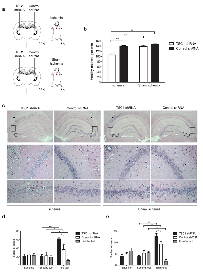
Figure 3. Resistance of CA3 neurons to ischemia is mediated by upregulation of hamartin in vivo.
(a) Design of experiments in which rats were stereotaxically injected unilaterally with a TSC1 shRNA vector in the dorsal CA3 pyramidal layer and subjected to either ischemia or sham ischemia (n=5). (b) Quantification of the number of neurons per mm in the dorsal CA3 pyramidal layer (one-way ANOVA with Bonferroni’s post hoc test, **p<0.01) from rats treated as in (a). (c) Representative hematoxylin and eosin stained hippocampal sections from rats treated as described in (a). Arrowheads show the needle tract. Scale bars: 1.00 mm (top row), 0.01 mm (middle and bottom rows). Boxed areas indicate the magnified CA3 regions shown in the bottom images. (d,e) Quantification of boxes crossed (d) and rears performed (e) in an open field test on rats bilaterally injected with TSC1 shRNA (n=6) or control shRNA (n=4) and subjected to ischemia (two-way ANOVA with Bonferroni’s post hoc test, *p<0.05, ***p<0.001). Open field test was carried out at three time-points: baseline, immediately before ischemia (second test) and 7 d after ischemia (third test). Data are mean ± S.E.M.

