
Figure 6.
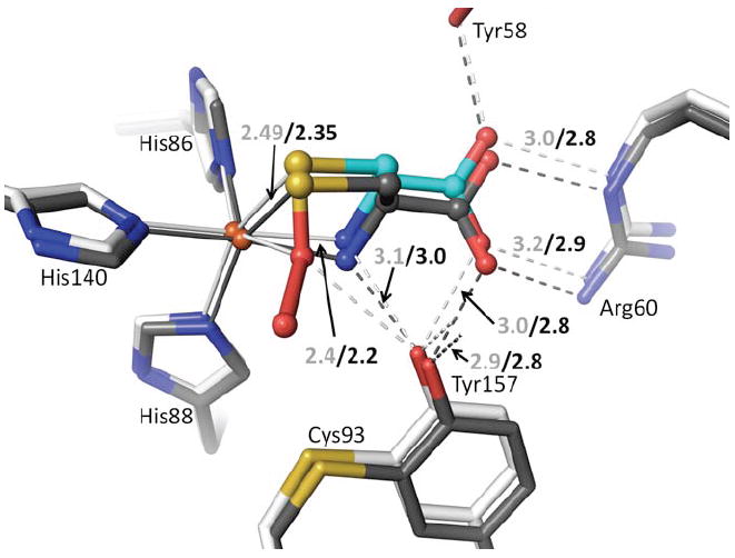
Active site differences between Cys-bound and Cys-persulfenate bound CDO. The persulfenate complex at pH=6.2 (cyan and white carbons) is overlaid on the Cys-bound complex pH=9.0 (dark carbons). Coordination and H-bond distances are given for the persulfenate complex (light text) and for the Cys-bound complex (black text). In the Cys-bound complex the Cys N- and Sγ-ligands are moved slightly to partly occupy the space left by the missing sixth ligand. This movement causes a slight shift in the whole Cys orientation and of the Cys-Tyr and Arg that interact with it.