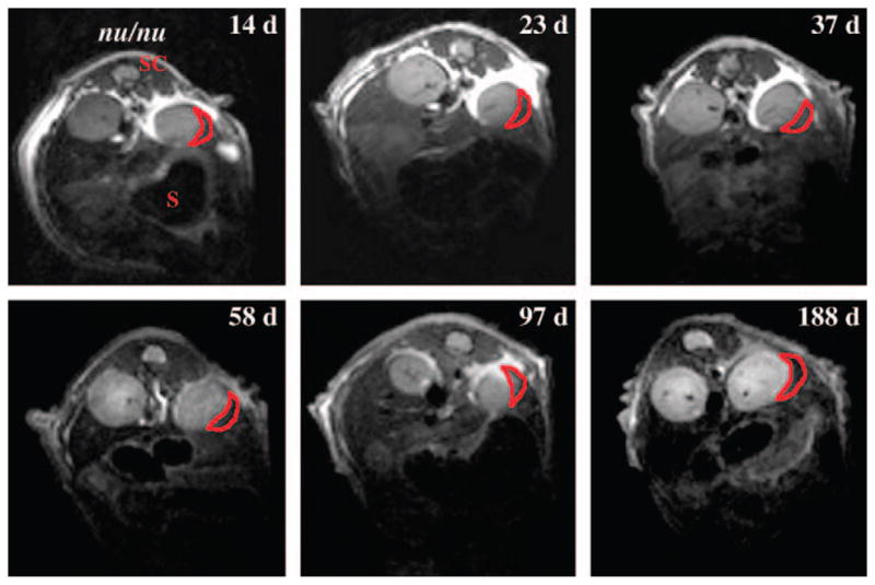
Fig. 1.

In vivo MRI of islet transplantation under the kidney capsule. Transverse T2-weighted magnetic resonance images of transplanted labeled and non-labelled human islets 14, 23, 37, 58, 97 and 188 days after transplantation under the kidney capsule in nude (nu/nu) mice. The dark area in the left kidney represents a labeled graft (red outline). No darkening was reported for the right kidney with unlabelled graft. S, stomach; SC, spinal cord. Reprinted with permission from Macmillan Publishers Ltd [52].
