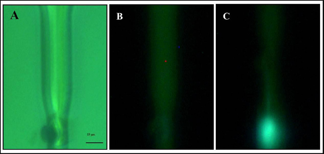
Figure 9.
Test of intracellular delivery. The ability of a jet to pierce a cell was tested firing a solution with a potassium indicator into a cell suspended in a potassium free buffer. A) Bright field picture of channel and cell. The cell is partially squeezed in a narrowing of the channel and is positioned in front of the micro nozzle B) Fluorescence picture just before firing a jet. C) Fluorescence image just after firing the jet. The indicator starts fluorescing after reacting with potassium present inside the cell. The fluorescence appears confined within the cell boundaries showing that the cell maintains its structure.

