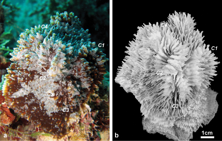
Figure 2.
Holotype of Echinophyllia tarae sp. n. (MNHN-IK.2012–8000) a the colony in situ before collection, and b the corallum after removal of the animal tissues. C1 to 4 indicate the position of same corallite (C) in the two images. Numbers are assigned in decreasing order of corallite size, C1 being the largest. Stippled line on the specimen in b shows the boundary of living tissue at the time of collection.

