Figure 2.
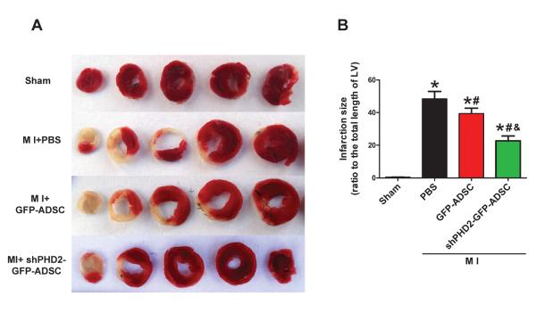
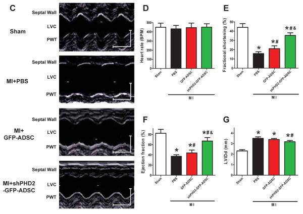
Transplantation of ADSCs with PHD2 silencing reduces infarct size and improves cardiac function in MI mice. A: Representative images of TTC-stained heart sections obtained from Sham, MI+PBS, MI+GFP-ADSCs, and MI+shPHD2-GFP-ADSCs groups at 4 weeks after transplantation. B: LV infarct sizes expressed as the ratio of the length of the infarct band to the total length of LV (N=14). C: Representative M-mode images of hearts with sham surgery or MI at 4 weeks after PBS, GFP-ADSCs and shPHD2-GFP-ADSCs injection. Scale bar: X axis: 0.1 second; Y axis: 0.2 cm. D: Heart rates were controlled to be similar in different groups. E–G: LV fraction shortening (E), LV ejection fraction (F) and diastolic left ventricle internal diameter (LVIDd, G) at 4 weeks after ADSCs transplantation. N=11. * p<0.05 vs. Sham; # p<0.05 vs. MI+PBS; & p<0.05 vs. MI+GFP-ADSCs.


