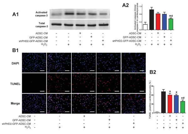
Figure 6.
The anti-apoptotic effect of ADSCs conditioned medium (CM) on cardiomyocytes subjected to oxidative injury. A: Representative blots (A1) and quantification (A2) of Western blotting analysis of activated caspase-3 of NRVMs treated with H2O2 (N=7). B: Representative images (B1) and quantification (B2) of TUNEL staining in cardiomyocytes subjected to H2O2 treatment (N=16). Cell nuclei were stained with DAPI (blue) and TUNEL+ nuclei were labeled with TMR-red. TUNEL positive rate= (TUNEL positive nuclei / DAPI positive nuclei)×100%. *p<0.05 vs. Control with H2O2; # p<0.05 vs. H2O2+GFP-ADSC-CM.

