
Figure 5.
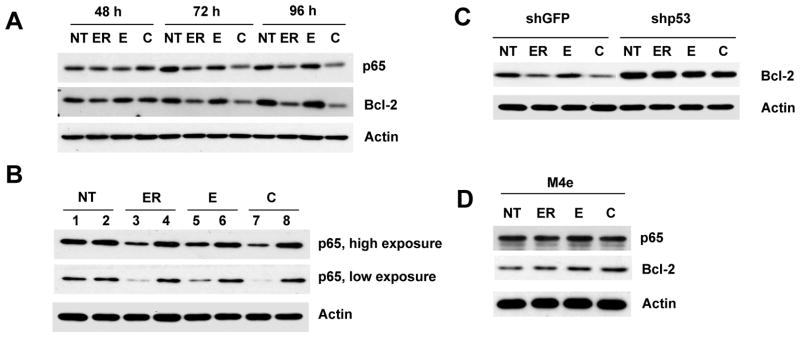
p53-dependent inhibition of NF-κB. (A) Tu686 cells were treated with 2 μM erlotinib, 30 μM EGCG and a combination of 2 μM of erlotinib and 30 μM EGCG for the indicated times. Expression of p65 and Bcl-2 was measured by immunoblotting. (B) shGFP- and shp53-transduced Tu686 cells were treated with 2 μM erlotinib, 30 μM EGCG and a combination of 2 μM of erlotinib and 30 μM EGCG for 72 h. Total cell lysates were immunoblotted with anti-p65. Lanes 1, 3, 5, 7 are shGFP and lanes 2, 4, 6, 8 are shp53. (C) Cells were treated as in Figure 5B and total cell lysates were used for the expression of Bcl-2. (D) M4e cells were treated with 0.5 μM erlotinib, 30 μM EGCG and a combination of 0.5 μM of erlotinib and 30 μM EGCG for 48 h and total cell lysates were immunoblotted with anti-p65 and anti-Bcl-2. Reproducibility of all results were confirmed by three independent experiments.