
Figure 4.
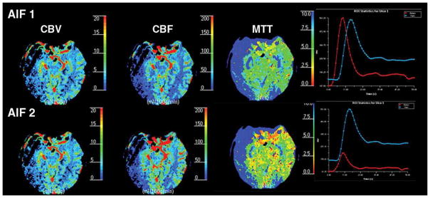
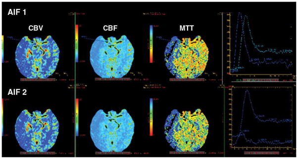
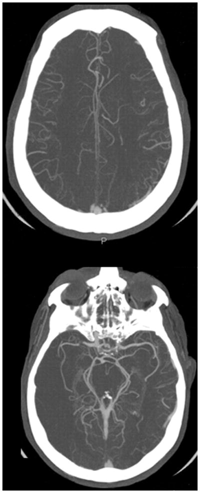
Figure 4A. 52-year-old man with acute right-sided middle cerebral artery infarction. CT perfusion color maps using software B (standard technique) obtained with arterial input function (AIF) 1 and AIF 2 in acute stroke patient with robust collateral circulation. No significant difference is observed in perfusion maps obtained with AIF 1 and AIF 2. Delay in contrast arrival and reduction in height of arterial time–attenuation curve are less prominent with AIF 2, and full width at half maximum was not affected.
Figure 4B. 52-year-old man with acute right-sided middle cerebral artery infarction. CT perfusion color maps using software A (delay-insensitive technique) obtained with AIF 1 and AIF 2 also show no significant difference.
Figure 4C. 52-year-old man with acute right-sided middle cerebral artery infarction. CT angiography source images show robust collateral circulation.