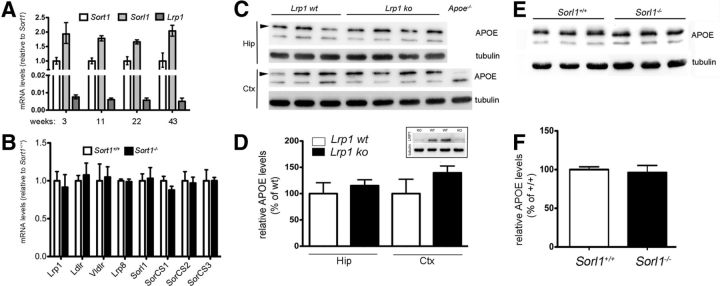
Figure 12.
Functional expression of APOE receptors in the murine brain. A, Quantitative RT-PCR of levels of Sort1, Sorl1, and Lrp1 transcripts in the cortex of wild-type mice of the indicated ages (n = 3 per group). B, Quantitative RT-PCR of levels of the indicated receptor transcripts in the cortex of wild-type and sortilin-deficient mice (n = 4 per group). C, D, Levels of APOE in hippocampus (Hip) and cortex (Ctx) of wild-type mice (Lrp1 wt) or animals with neuron-specific Lrp1 gene defect (Lrp1 ko) were determined by Western blotting (C) and quantified by densitometric scanning of the respective blots (n = 3–4) (D). Detection of tubulin served as loading control. Extracts from mice genetically deficient for APOE (Apoe−/−) were used as negative control for specificity of the APOE immunoreactivity (APOE indicated by arrowheads). The inset in D demonstrates robust expression of LRP1 in brain extracts of wild-type mice but not of animals with neuron-specific Lrp1 deletion. E, F, Levels of APOE in cortex of wild-type (Sorl1+/+) or SORLA-deficient (Sorl1−/−) mice were determined by Western blotting (E) and quantified by densitometric scanning of the respective blots (n = 6) (F). Detection of tubulin served as loading control.

