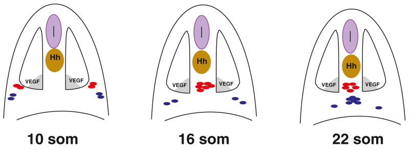
Figure 7. A Model for Arterial-Venous Differentiation.
At the 10-somite stage endothelial progenitor cells are positioned within the LPM in two bilateral lines, a medial (red) and lateral line (blue). After the 10-somite stage, medial angioblasts migrate intersomitically to the midline directly to the dorsal position where they differentiate as arterial cells. Shortly after the 15-somite stage, the lateral angioblasts migrate to the ventral position at the midline where they differentiate as venous endothelial cells. Hh, expressed in the notochord; VegfA, expressed in the ventral somites function as morphogens and are important for specifying arterial fate in the medial angioblasts.

