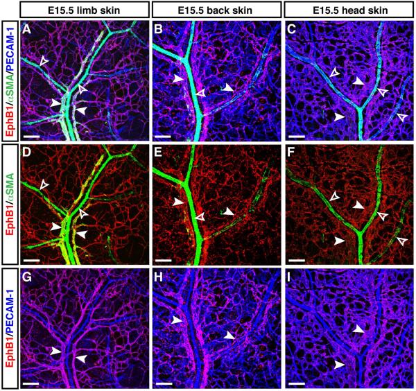
Figure 5. Venous expression of EphB1 in embryonic limb, back and head skin vasculatures.
Whole-mount triple immunofluorescence analysis of forelimb skin (A, D, G), back skin (B, E, H), and head skin (C, F, I) of E15.5 embryos was performed with antibodies to the vascular smooth muscle cell (VSMC) marker αSMA (A-F, green) in combination with EphB1 (A-I, red) and PECAM-1 (A-C, G-I, blue). At E15.5, arterial branches are densely covered with αSMA+ VSMCs (A-F, open arrowheads), whereas less or no VSMC coverage occurs in venous branches (A-F, arrowheads). EphB1 expression was detectable in venous vasculature in limb, back and head skin vasculatures. Scale bars are 100 μm.

