
Figure 3.
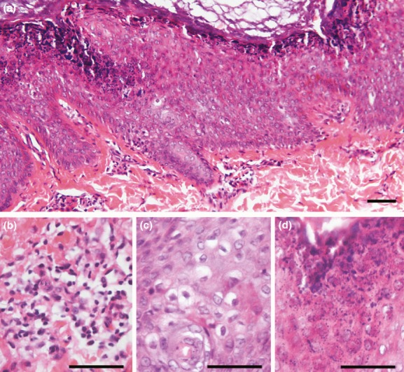
Histopathological findings. (a) Histology of cutaneous forearm samples indicate compact hyperkeratosis. (b) Presence of dermal inflammatory infiltrates. (c) Acanthosis with vacuolar degeneration of suprabasal keratinocytes. (d) Expanded granular layer with increased number of coarse keratohyaline granules. Scale bar = 50 μm.