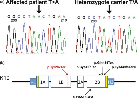
Figure 4.

Mutation localization in the context of K10 protein structure. (a) Exon 3 mutation showing homozygous T→A mutation (p.Tyr282Ter.) and a heterozygous carrier. (b) Schematic representation of K10 protein structure. Previous recessive epidermolytic ichthyosis (EI) mutations in the 2B domain are denoted in black. p.Tyr282Ter. (red) is located in the 1B domain of the protein.
