
Figure 5.
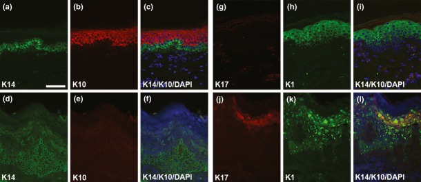
Keratin expression. Immunofluorescence staining of control (foreskin, a–c, g–i) and affected samples epidermolytic ichthyosis (EI, d–f, j–l) for K14 (a, d), K10 (b, e), K17 (g, j), and K1 (h, k). In EI samples, note the absence of K10. Ectopic expression of K14 in the suprabasal layers and expression of K17 in the interfollicular epidermis may compensate for the loss of K10. Also, note the clumpy appearance of K1 in the upper spinous and granular layers. Scale bar = 50 μm.