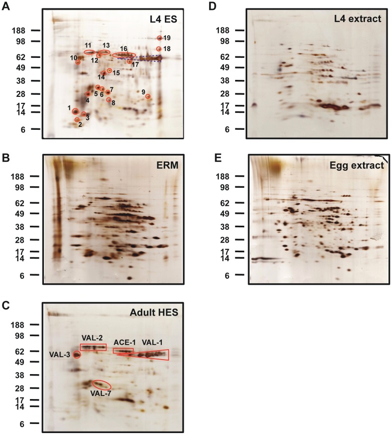
Figure 1. 2-D gel electrophoresis of ES released material and somatic extracts from different stages of H.polygyrus, visualised by silver staining.
A. L4 ES. Identities of numbered spots are given in Table 1. B. Egg released material (ERM). C. Adult HES. Solid red boxes correspond to the indicated protein products. D. L4 somatic extract. E. Egg somatic extract. Positions of molecular weight markers (kDa) are indicated.

