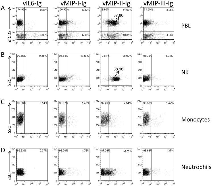
Figure 1. vMIP-II-Ig binds PBLs and naïve NK cells.
Freshly isolated PBLs (A), freshly isolated naïve NK cells (B), Monocytes (C) and Neutrophils (D), (the various cell types are indicated in the right of the figure), were stained with different KSHV chemokines fused to human IgG1: vIL6-Ig, vMIP-I-Ig, vMIP-II-Ig or vMIP-III-Ig (X axis, indicated in the top of the figure). PBLs were double stained with the indicated chemokines fused to IgG and with anti-CD3. The percentages of various populations are indicated inside the dot plot. The Median Fluorescence Intensity (MFI) of the vMIP-II-Ig staining of CD3+ cells (A) and of NK cells (B) is indicated and is marked by an arrow. Figure shows one representative staining out of more than 3 performed.

