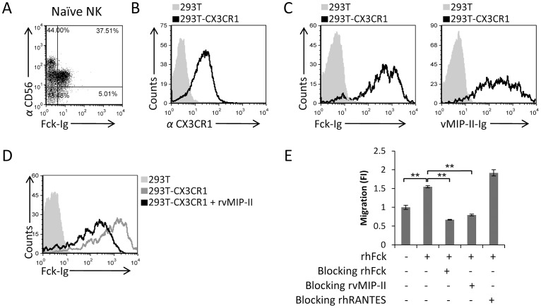
Figure 5. vMIP-II blocks the migration of freshly isolated naïve NK cells to Fractalkine.
(A) Freshly isolated naïve NK cells were double stained with Fck-Ig and with anti-CD56 mAb. The percentages of the various populations are indicated in the figure. (B) CX3CR1 expression on the transfectant 293T-CX3CR1 cells (black open histogram) or on 293T parental cells (filled grey histogram). (C) Binding of Fck-Ig (left histogram) or vMIP-II-Ig (right histogram) to 293T-CX3CR1 transfectant (black open histogram) or to the 293T parental cells (filled grey histogram). (D) 293T-CX3CR1 cells were incubated with (black empty histogram) and without (dark gray empty histogram) rvMIP-II for 1 hour in 4°C and then stained with Fck-Ig. The light gray filled histogram is the staining of Fck-Ig on the 293T parental cells. (E) Freshly isolated naïve NK cells were incubated at 4°C for 1 hour with and without the proteins indicated in the x axis. RhFck was placed in the bottom chamber and the numbers of migrated cells was determined by FACS following 3 hours incubation at 37°C. The migration of the NK cells without the appropriate chemokine was set as 1 and the results are presented as fold increase (FI). **P<0.01. NS - not significant. Figure shows one representative experiment out of four performed.

