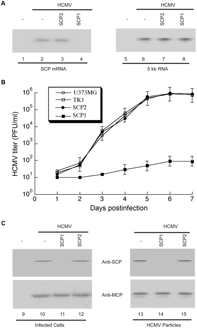
Figure 4. Ribozyme-mediated inhibition of HCMV SCP expression and viral growth.
(A) Northern analysis of HCMV mRNAs in infected cells. RNA samples were isolated from parental U373MG cells (lanes 1, 2, 5 and 6) or M1GS-expressing cells (lanes 3, 4, 7 and 8) that were either mock-infected (lanes 1 and 5) or infected with HCMV (MOI = 0.5–1; all other lanes) for 48 h, separated by denaturing gels, and transferred to membranes. Membranes were hybridized with radiolabled probes containing the sequence of HCMV SCP mRNA (lanes 1–4) or IE 5 kb RNA (lanes 5–8). SCP1, ribozyme targeting HCMV SCP mRNA for degradation; SCP2, control ribozyme that binds but cannot degrade HCMV SCP mRNA. (B) Growth of HCMV in U373MG cells and cell lines expressing M1GS RNA. Cells (5×10⁵) were infected with HCMV at MOI = 3. Values are means derived from triplicate experiments. Standard deviation is indicated by error bars. TK1, control ribozyme targeting HSV-1 thymidine kinase mRNA. (C) Western analysis of HCMV SCP and MCP proteins. Protein samples were either isolated from the parental U373MG cells or ribozyme-expressing cells (Infected cells, lanes 9–12) or from viral particle preparations purified from these cells (HCMV particles, lanes 13–15), separated in SDS-polyacrylamide gels, transferred to membranes, and reacted with antibodies against HCMV SCP (anti-SCP) and MCP (anti-MCP) [24].

