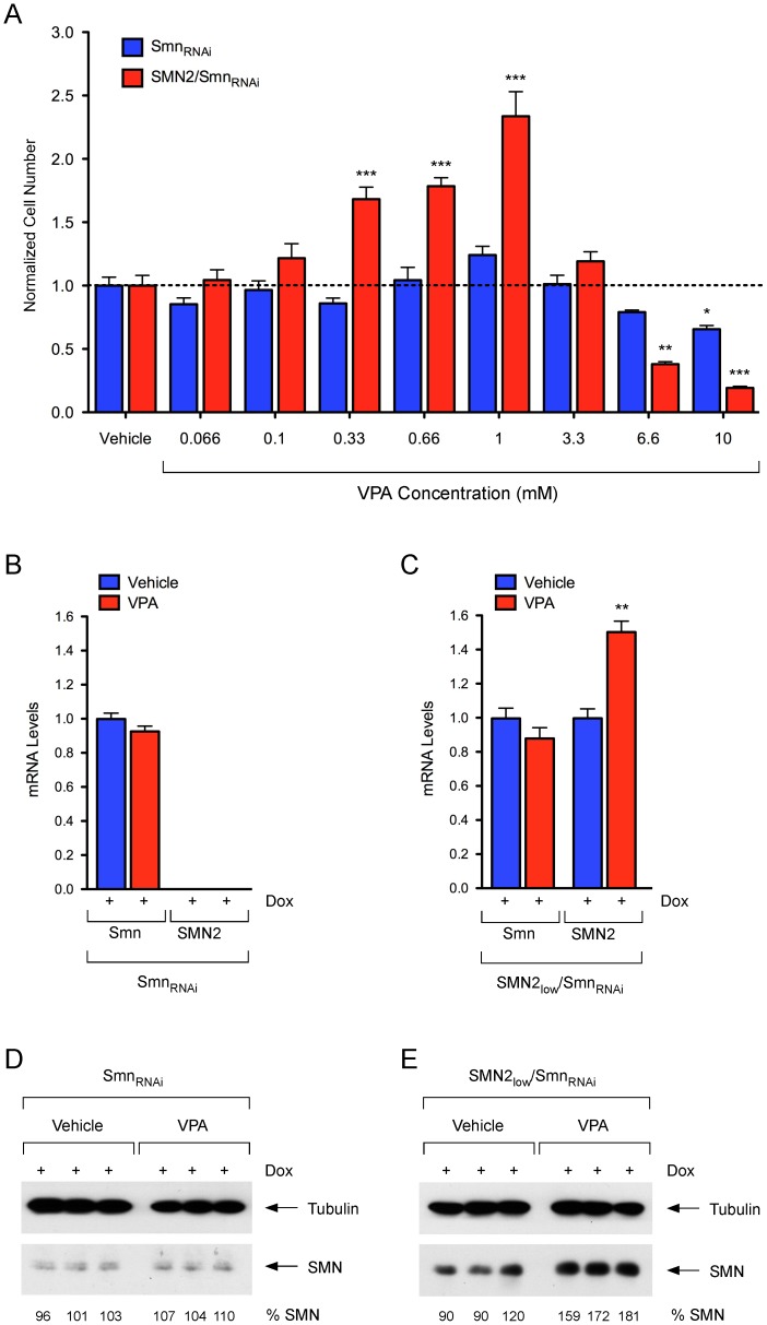
Figure 8. Pharmacological modulation of SMN-dependent proliferation in NIH3T3 cells.
(A) Dose-response analysis of VPA treatment on cell proliferation of SMN-deficient NIH3T3-SmnRNAi and NIH3T3-SMN2low/SmnRNAi cells. In these experiments, NIH3T3 cells were cultured for 5 days with Dox prior to seeding in six replicate wells of a 96-well plate in the presence of Dox. Vehicle or increasing amounts of VPA were added 4 hours later. For each group and treatment, cell number was determined at 5 days post-plating and normalized to that of the corresponding vehicle-treated cells. Data are represented as mean and SEM (n = 6; * = p<0.05; ** = p<0.01; *** = p<0.001; one-way ANOVA). (B–C) RT-qPCR analysis of mouse Smn and human SMN2 mRNA levels in NIH3T3-SmnRNAi (B) and NIH3T3-SMN2low/SmnRNAi (C) cells cultured with Dox for 3 days and then in the presence of either water (Vehicle) or 1 mM VPA for additional 4 days. Data from triplicate RT-qPCR experiments normalized to Gapdh mRNA are represented as mean and SEM (** = p<0.01; t-test). (D–E) Western blot analysis of SMN protein levels in NIH3T3-SmnRNAi (D) and NIH3T3-SMN2low/SmnRNAi (E) cells cultured with Dox for 3 days and then in the presence of water (Vehicle) or 1 mM VPA for additional 4 days. Blots were probed with an antibody that recognizes both mouse and human SMN proteins. SMN levels in VPA-treated relative to vehicle-treated cells are shown at the bottom.

