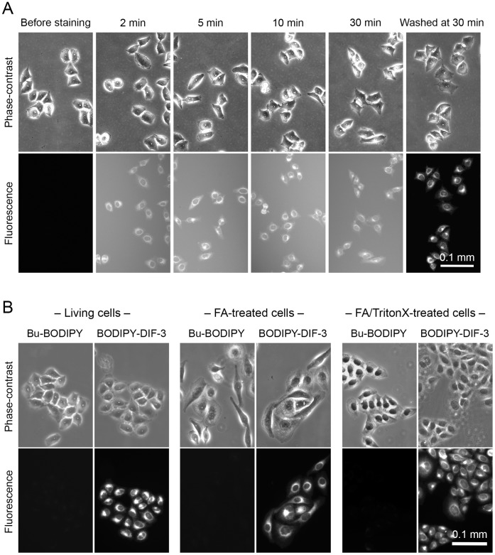
Figure 2. Membrane permeability and localization of BODIPY-DIF-3 and Bu-BODIPY in living, formalin-fixed, and detergent-treated HeLa cells.
(A) Cells were incubated with 20 µM BODIPY-DIF-3 and observed microscopically at the indicated time points. After 30 min of incubation, cells were washed free of the additive and observed again. BODIPY-DIF-3 rapidly penetrated the cell membrane within 2 min of exposure, reaching a maximal level within 30 min. (B) Living cells, formalin (FA)-fixed cells, and FA-fixed and TritonX-100-treated cells were incubated for 0.5 h with 20 µM of Bu-BODIPY or BODIPY-DIF-3, washed free of the additives, and observed microscopically. All cell samples stained well with BODIPY-DIF-3 but not with Bu-BODIPY.

