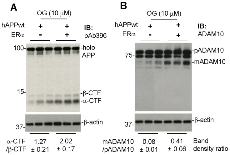
Figure 4. OG fails to promote α-secretase cleavage of APP in estrogen receptor α (ERα) deficient cells.
We cultured primary cortical neuronal cells from brain tissues of one-day-old ERα deficient and intact mice. One week after the primary culture, these neuronal cells were transiently transfected with human wild-type APP695 plasmid DNA (hAPPwt) with Lipofectiamine™ LTX Reagent (as detailed in the method section). Twenty four hours later, these neuronal cells were treated with OG at 10 µM for 12 hours. Cell lysates were then prepared and subjected to IB analyses for α- and β-CTF (A) using pAb396 and ADAM10 maturation (B) using an anti-carboxyl-terminal ADAM10 antibody (ADAM10). Most notably, OG’s promotion of anti-amyloidogenic APP processing was significantly attenuated in the ERα null mouse-derived primary neuronal cells overexpressing hAPPwt. Human APP expression was examined after transient transfection by IB analysis using an anti-Aβ1–17 antibody (6E10) (data not shown). As shown below each IB, densitometry analysis shows the band density ratio (mean ± SD) of α-CTF to β-CTF and mature ADAM10 (mADAM10) to pre-mature ADAM10 (pADAM10).

