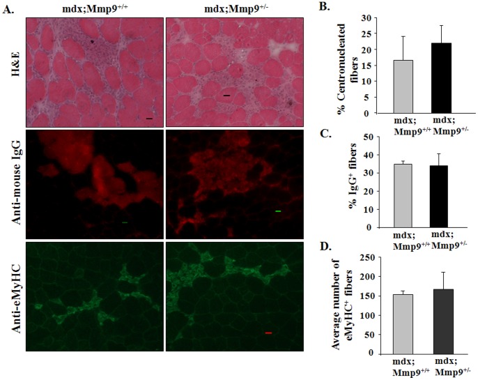
Figure 1. MMP-9 is not involved in the initial onset of muscle pathology in mdx mice.
(A) Representative photomicrographs of GA muscle sections of 4-week old mdx;Mmp9+/+ and mdx;Mmp9+/− mice displaying hallmarks of mdx pathology. Area under necrosis and centronucleated myofibers evaluated by H&E staining (top panel). Muscle sections were immunostained with Cy3-labeled goat anti-mouse IgG to detect permeable/damaged fibers (middle panel). Formation of new myofibers was assessed by staining with embryonic myosin heavy chain (eMyHC) antibody (lower panel). Scale bar: 20 µm. Quantification of (B) percentage of centronucleated fibers; (C) Cy3-labelled IgG filled fibers per field; and (D) number of eMyHC-positive fibers per field of mdx;Mmp9+/+ and mdx,MMP9+/− mice. N = 4 in each group. Error bars represent SD.

