
Figure 1. 3 mo post aerosol infection with ~100 M. tuberculosis H37Rv CFU I/St mice display substantially more severe infectious course compared to B6 mice.
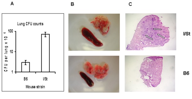
(A) -1 log difference in lung CFU counts (N=5, P<0.001, ANOVA, 1 experiment of 3 similar); (B) – more prominent gross pathology of the lung and greater splenomegaly; (C) – granulomata with necrotizing centers (arrows) are present in the lungs of I/St but not of B6 mice (X25).