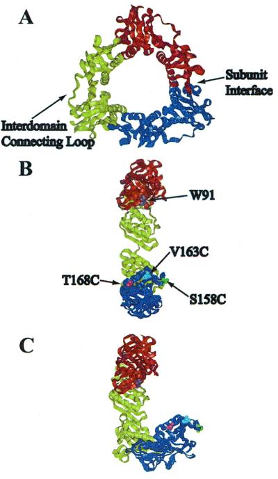
Figure 1.
(A) X-ray crystal structure of gp45 showing the interdomain connecting loop and the subunit interface. (B) In-plane model of opening of gp45 showing the location of the mutations: V163C in blue, S158C in green, and T168C in pink, with the donor tryptophan in orange. Each mutation is shown only once for clarity. (C) Out-of-plane model of opening.

