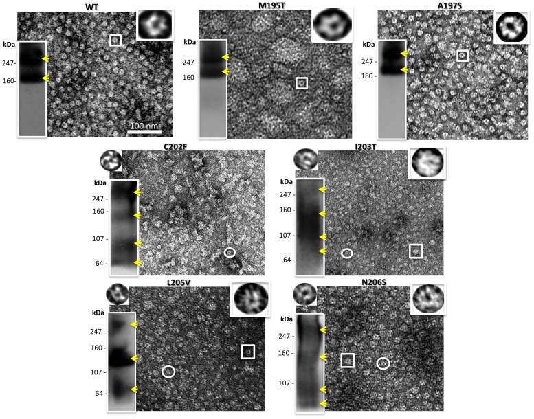
Figure 4. Analysis of hemichannel stability for gap junction forming mutants.
Shown here are a representative electron micrograph and Blue Native westerns (bottom left hand inset) of purified hemichannels for each mutant purified from an Sf9/baculovirus expression system. The yellow arrowheads point to the recognizable bands. The insets on the top right are 6 fold enlargements of a normal hemichannel (the expected hexamer) indicated by the box in the micrograph, while the insets on the top left are 6 fold enlargements of the small oligomers visible only in unstable mutants and indicated by the circle in these micrographs.

