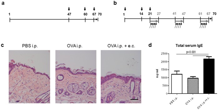
Figure 1. Sensitization protocol, histological analysis, and IgE serum levels.
(a) Mice were sensitized i.p. with 10 µg OVA adsorbed to 1.5 mg Al(OH)3 or with phosphate-buffered saline (PBS; control) on days 47, 60 and 67 (black arrows). (b) A third group of mice was sensitized i.p. with 10 µg OVA adsorbed to 1.5 mg Al(OH)3 on days 1, 14 and 21 (black arrows) followed by e.c. OVA exposure for three 1-week periods (grey arrows) separated by 2-weeks intervals. Each mouse received a weekly e.c. dose of 100 µg OVA adsorbed to 1.5 mg Al(OH)3 in 100 µl PBS on shaved back skin divided into four applications of 25 µl every other day of one week (black angular arrows). Three days after the last treatment (day 70), mice were sacrificed and skin and serum samples were collected. (c) Hematoxylin and Eosin staining of five-micrometer skin sections obtained from treated dorsal skin sites. Images were taken at ×10 magnification (scale bar = 50 µm). (d) Total IgE levels were determined in the serum of mice treated systemically with or without additional topical sensitization with OVA. Data are presented as mean values ± SEM of three independent measurements with triplicate determination of n = 8 mice/group. Statistical significance (p) is based on one-way ANOVA followed by Tukey’s multiple comparison test.

