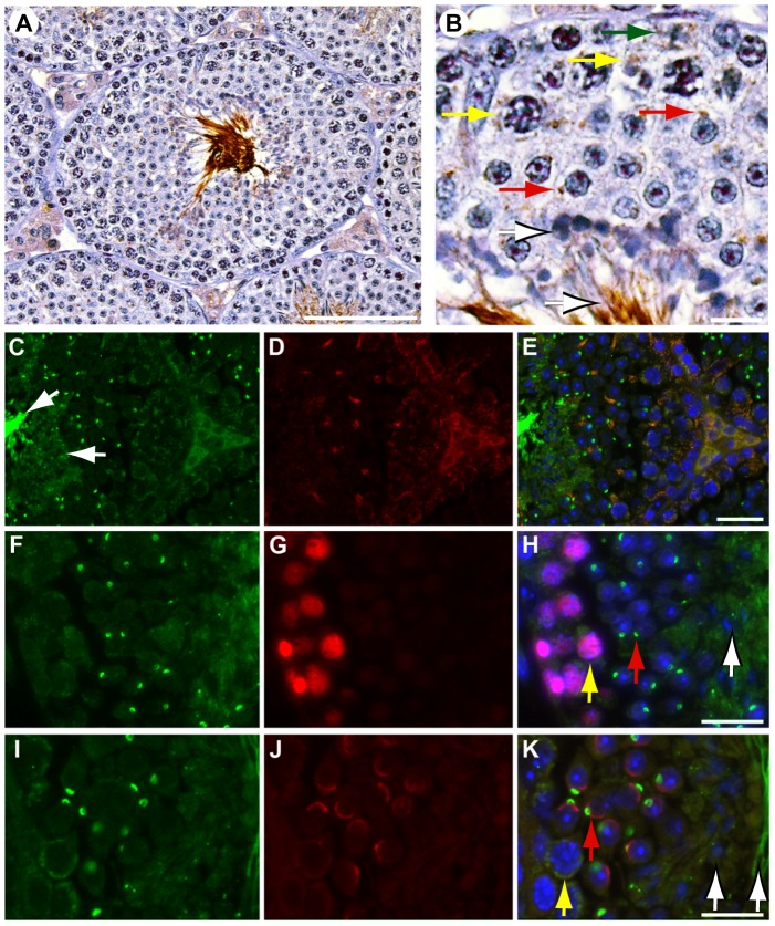
Figure 1. SLIRP is expressed in the mouse testis.
(A & B) Detection of SLIRP by immunohistochemistry in Leydig cells, spermatogonia (green arrow), early spermatocytes (yellow arrows), round spermatids (red arrows), elongate spermatids and sperm lining the lumen (white arrows). (C, F, I) Immunofluorescent staining for SLIRP (green), (D) Hsp60 (red), PCNA (G, red) and SP56 (J, red) overlayed in E, H & K respectively. Yellow indicates colocalization in overlayed panels. Nuclear DAPI staining in blue E, H & K. White bars: A, 80 µm, B, H & K, 10 µm, E, 20 µm.

