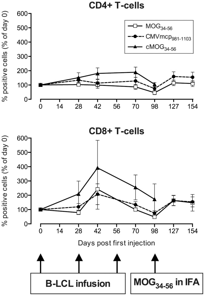
Figure 4. In vivo expansion of T-cells.

Percentages of CD4+ T-cells and CD8+ T-cells in PBMC, expressed as a percentage of day 0, demonstrate that mainly the CD8+ subpopulation is expanded after the infusion of B-LCL, which is consistent with the data in Figure 1. Percentages of CD4+ T-cells on day 0 were: 35.7±11.2; 36.8±9.8 and 29.5±19% for groups A, B and C respectively, which is not significantly different from each other (P = 0.4677). Percentages of CD8+ T-cells on day 0 were: 18.4±3.5; 21.2±5.2 and 10.2±2.5% for groups A, B and C respectively (P = 0.1451). Consistent with our published data in rhesus monkeys [17] an expansion of both CD4+ and CD8+ T-cells was observed after a booster with MOG34–56 in IFA.
