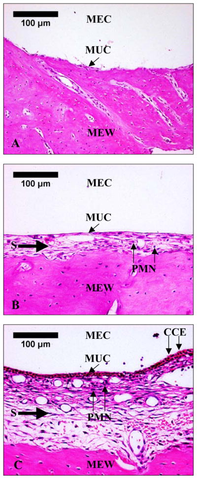
Figure 3.

Histological photographs of middle ear roof mucosa of (A) control ear, (B) OME ear, and (C) AOM ear 3 days after inoculation. MEC, middle ear cavity; MUC, middle ear mucosa; MEW, middle ear bony wall; S, submucosal layer; PMN, polymorphonuclear leukocyte; CCE, ciliated columnar epithelium.
