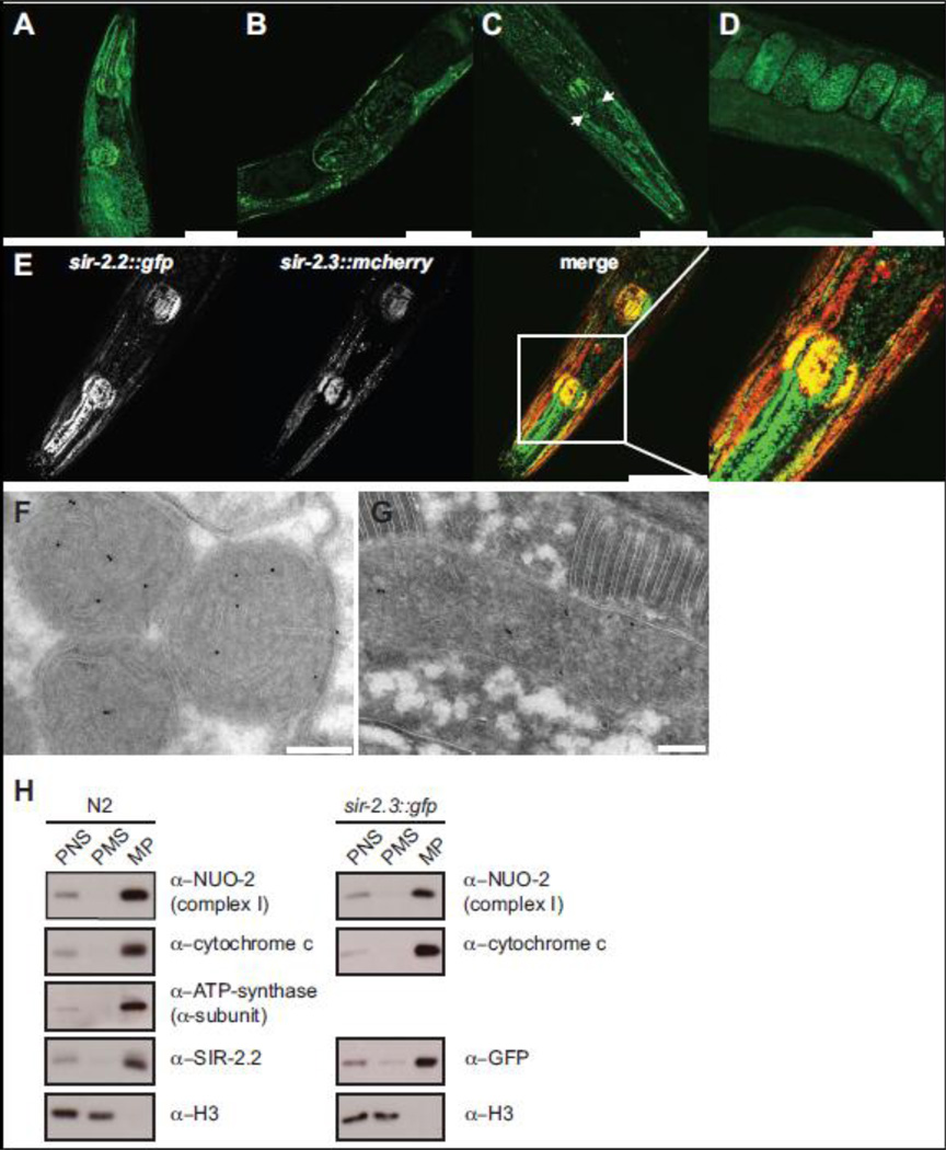
Fig. 1. C. elegans SIR-2.2 and SIR-2.3 are mitochondrial proteins.
(A) and (B) representative fluorescent confocal images of sir-2.2::gfp transgenic worms showing SIR-2.2::GFP expression in the head region and gonad of adult C. elegans. (C) and (D) SIR-2.3::GFP expression and localization in the head and gonad of adult worms. Arrows point at unidentified cells in the head. (E) Expression and localization of SIR-2.2::GFP and SIR-2.3::mCherry in the head of double transgenic sir-2.2::gfp; sir-2.3::mcherry worms. Scale bars represent a magnification of 50 µm. (F) Immunogold labelling of wild type N2 worm using the anti-SIR-2.2 antibody. (G). Representative cryo section of sir-2.3::gfp transgenic worm stained with anti-GFP antibody. Immunocomplexes were visualized with protein A-conjugated gold beads (10 nm). High-density black dots indicate the localization of SIR-2.2 and SIR-2.3 to mitochondria. Scale bars represent a magnification of 200 nm. (H) Wild type N2 worms and sir-2.3::gfp transgenic worms were homogenized and subjected to subcellular fractionation by differential centrifugation. Equal protein amounts of the post nuclear supernatant (PNS), mitochondrial pellet (MP) and post mitochondrial supernatant (PMS) were analyzed by SDS-PAGE and Western blotting, using the indicated antibodies.

