
Figure 3. Acute increases in synaptic GluK2 do not involve insertion of de novo KARs.
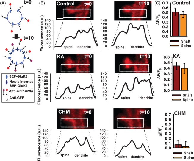
A) Schematic of protocol (see Materials and Methods for details). SEP-GluK2 inserted during the 10-min incubation interval illustrated by difference between the labelling at t = 0 and t = 10 min. Receptors previously surface expressed bound to unlabelled antibody and recycled back to the surface are not detected in this assay. Newly labelled receptors in each incubation are shown in red. Thus, receptors labelled at t = 0 are indicated as purple at t = 10, and the newly labelled receptors in red. B) Profiles of SEP-GluK2 insertion into spine and shaft. Images show SEP-GluK2 labelled with anti-GFP-Alexa 594 antibody at t = 0 and t = 10 in control cells, after kainate application or following 2-h incubation with cycloheximide. Note that in these experiments SEP is used as an extracellular epitope tag and only Alexa 594 fluorescence is shown and quantified. Scale bar 1 µm, n = 22–26 pairs spine/shaft per neuron from 10 to 12 neurons. C) Histograms showing the rate of insertion (calculated as ΔF/F0) in the spine and shaft of control cells (upper panel), kainate-treated cells (middle panel) or incubated with cycloheximide (bottom panel).