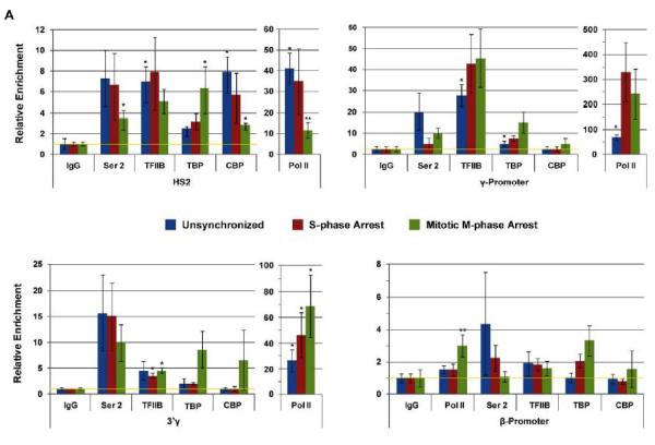
Figure 2.
Analysis of Pol II and transcription factor binding at the human β-globin gene locus in unsynchronized, synchronized (at the G1/S-border), and M-phase arrested cells. K562 cells were subjected to double thymidine block at G1/S-phase and nocodazole mediated arrest in early M-phase. Aliquots of the cells were taken from unsynchronized, G1/S-phase synchronized (indicated as S-phase arrest) and M-phase arrested (indicated as Mitotic arrest) cultures. The cells were subjected to ChIP assays using antibodies specific for Pol II, Pol II phosphorylated at serine 2 (indicated as Ser 2), TFII B, TBP, and CBP. Precipitation with IgG antibodies served as negative controls. The DNA was isolated from the immunoprecipitates and subjected to qPCR using primers specific for LCR element HS2, the Gγ-promoter region, the Aγ-3'end, and the β-globin promoter region. The data represent means +/− standard errors of the means of three independent experiments with PCR performed in duplicate. The data are shown relative to the IgG control, set at 1. The yellow line indicates IgG background levels.

