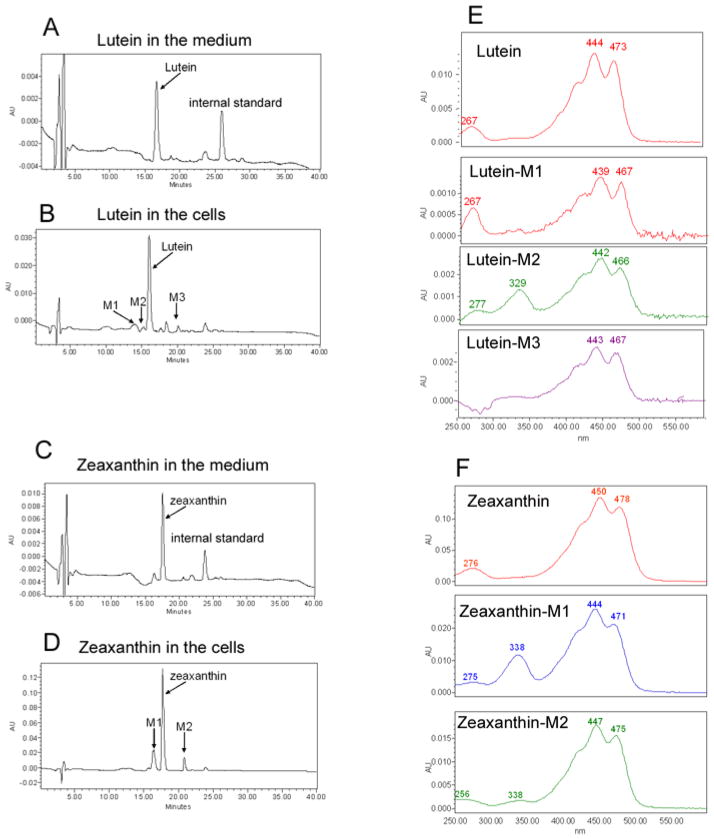
Fig. 1. Cultured RPE cells actively take up lutein and zeaxanthin.
Confluent ARPE-19 cells were cultured in media containing 1 or 10 μM of lutein or zeaxanthin for 3 days. The medium was removed and the cells were washed three times with medium containing 10% bovine serum. Then the cells were collected. Levels of lutein and zeaxanthin in the medium and in the cells were determined by reversed phase HPLC with a C30 column. This figure showed the chromatograms (monitored at 450 nm) and spectra of lutein and zeaxanthin in the medium and cells upon supplementation with 10 μM lutein or zeaxanthin for 3 days. Panel A: chromatogram of lutein in the medium; panel B: chromatogram of lutein and its putative metabolites in cells; panel C: chromatogram of zeaxanthin in the medium; panel D: chromatogram of zeaxanthin and its metabolites in cells; panel E: spectra of lutein and its metabolites in cells; panel F: spectra of zeaxanthin and its putative metabolites in cells.

