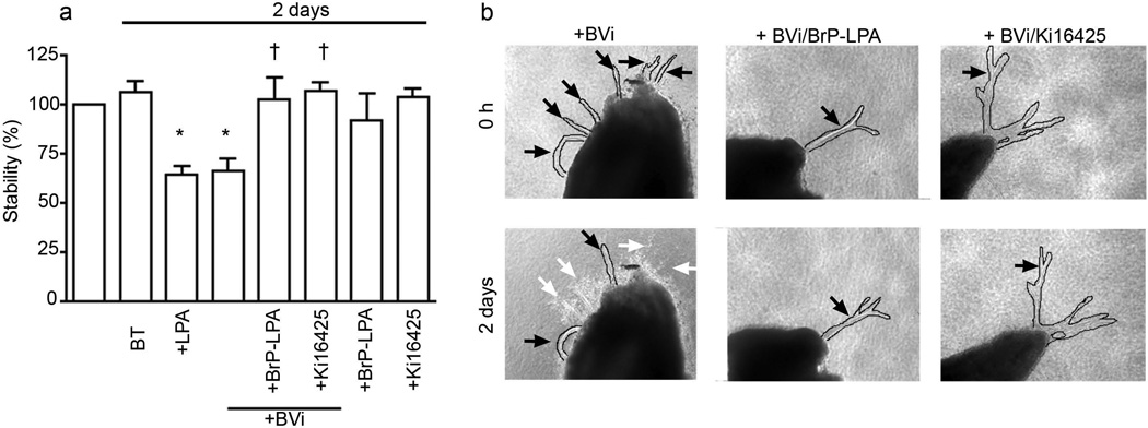
Fig. 1.
Regression of retinal explant vessels was induced by BVi in an LPA-receptor-dependent manner. Retinas were subjected to a retinal explant assay as described in the methods section. Lumenised vessels composed of endothelial cells developed in 2 weeks [26]. LPA (1 µmol/l) or BVi was added for 2 days; the treatments were renewed every 12 h. The vessel length was measured before and after the indicated treatments. Data shown are the means±SEM of four independent experiments. (a) Treatment with BVi induced regression comparable with that mediated by LPA. Addition of the LPA receptor inhibitors BrP-LPA (10 µmol/l) or Ki16425 (5 µmol/l) in combination with BVi inhibited regression. Addition of the inhibitors alone had no effect. The percentage stability is the ratio of vessel lengths before and after treatment. *p<0.05 vs explants treated with buffer; †p<0.05 vs explants treated with BVi; BT, buffer-treated. (b) Representative photographs of vessels treated with BVi alone or BVi treatment in the presence of BrP-LPA or Ki16425. Regressing vessels are indicated with white arrows. Stable vessels are indicated with black arrows. All vessels were digitally outlined to facilitate their visualisation (solid lines). Magnification is ×100 for all images

