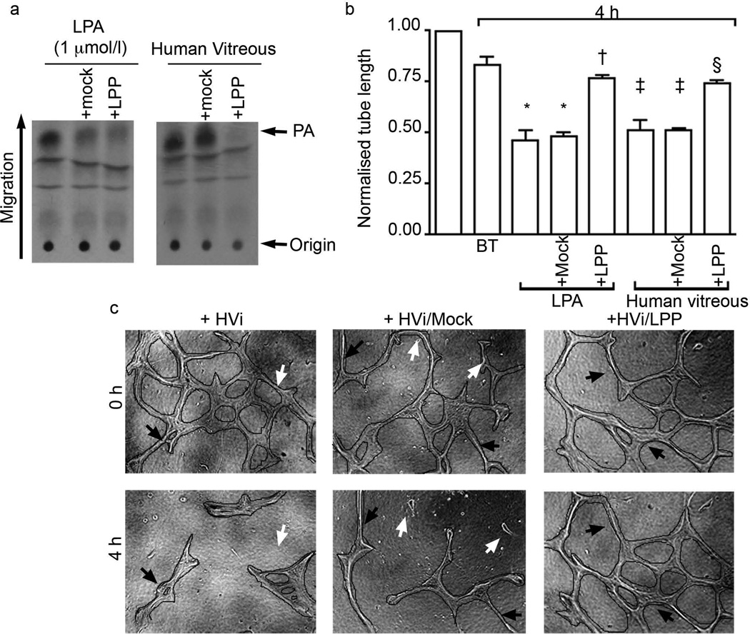
Fig. 3.
LPA was present in HVi and was critical for its vascular regression activity. (a) LPA was detected in HVi from patients with non-angiogenic retinal disease (as described in Table 1). (b,c) Control or HVi that was cleared of LPA was tested for its ability to induce regression of tubes as described in the legend to Fig. 2. Similarly to BVi, HVi contained LPA, which was an essential component of vitreous regression activity. Normalised tube length is the ratio of final and initial tube lengths. *p<0.05 compared with buffer-treated (BT) tubes; †p<0.05 compared with tubes treated with LPA or mock treatment; ‡p<0.05 compared with buffer-treated tubes; §p<0.05 compared with HVi- and mock-treated tubes

