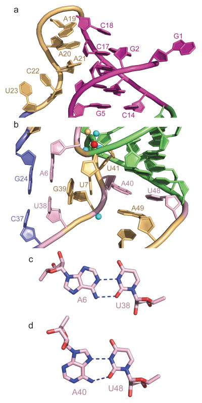
Figure 2. Details of long-range interactions within the structure of the T. petrophila fluoride riboswitch in the ligand bound state.
a, An expanded region of the structure highlighting pseudoknot formation involving residues G2-G3-C4-G5 of the 5′-overhang segment and residues C14-G15-C16-C17 of the large internal loop (in magenta), as well as continuous stacking within the A19-A20-A21 and C22-U23 steps. b, An expanded region of the structure highlighting long-range single-base pseudo-knot like pairing between A6 and U38, and between A40 and U48. Note the mutual interdigitation between unpaired U7 and G39 that contribute to formation of the junctional architecture. c, Long-range reversed Watson-Crick A6•U38 pair formation. d, Long-range reversed Hoogsteen A40•U48 pair formation.

