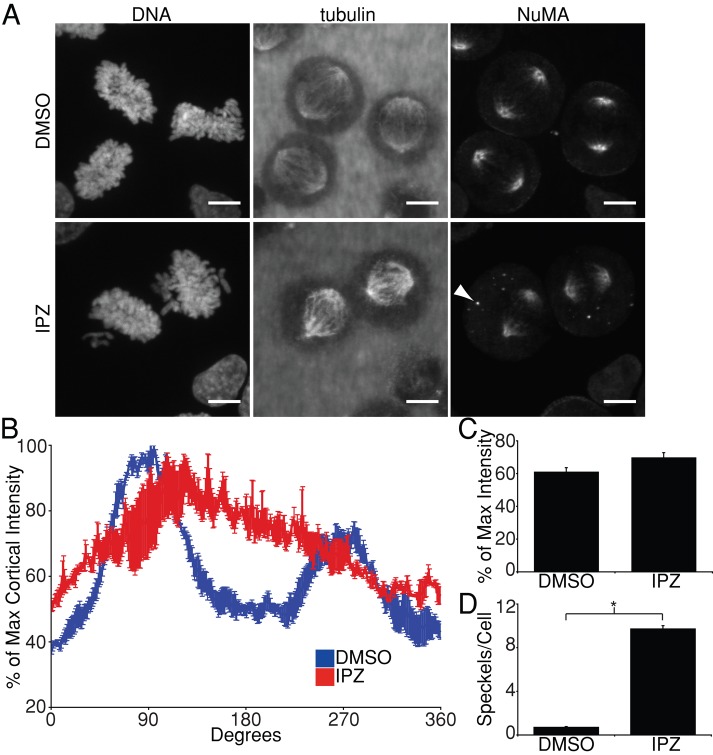
FIGURE 3:
Importazole impairs localization of cortical NuMA. (A) DNA, tubulin, and NuMA localization in synchronized mitotic HeLa cells treated with DMSO or 40 μM importazole for 1 h. NuMA in DMSO-treated cells localized to spindle poles, as well as to two arcs along the cortex in line with the long axis of the mitotic spindle, similar to the cortical staining pattern of LGN. NuMA's cortical localization was disrupted in importazole-treated cells. Cytoplasmic NuMA foci also appeared in importazole-treated cells (arrowhead). (B) Quantification of cortical NuMA staining in synchronized metaphase HeLa cells treated with DMSO or 40 μM importazole was performed as described in Figure 2B. (C) Mean cortical NuMA fluorescence as percentage of maximum intensity in DMSO and importazole-treated metaphase cells. (D) Quantification of the mean number of cytoplasmic NuMA foci observed per cell in DMSO- and importazole-treated cells. Scale bars, 10 μm. For B and C, n = 3, and 40 cells were measured per condition; for D, n = 3, and 20 cells were measured per condition. Bars, SE. Asterisk denotes statistical significance (p < 0.001).

