Figure 2.
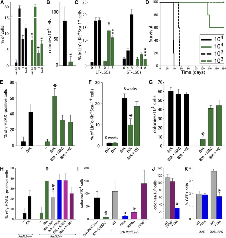
RAD52 DNA binding plays a critical role in BCR-ABL1-mediated leukemogenesis by preventing the accumulation of ROS-induced lethal DSBs. (A-D) BCR-ABL1 Rad52+/+ (black bars) and BCR-ABL1 Rad52−/− (green bars) murine bone marrow cells were analyzed for (A) cell cycle progression (*P < .05 in comparison with corresponding +/+ cells); (B) clonogenic activity (*P = .008); (C) frequency of long-term leukemia stem cells (LT-LSCs) and short-term leukemia stem cells (ST-LSCs) at 0, 4, and 8 weeks after BCR-ABL1 expression (*P = .04; **P = .001; ***P < .001 in comparison with the corresponding +/+ subpopulation); and (D) leukemia induction in SCID mice (5-6 mice/group). (E-G) BCR-ABL1-positive (B/A) and nontransfected (−) Rad52−/− cells (green bars) and Rad52+/+ counterparts (black bars) were incubated with N-acetyl-cysteine (NAC) and vitamin E (VE) when indicated. (E) Percentage of Lin−c-Kit+Sca-1+ cells with more than 20 γ-H2AX foci; *P < .01 in comparison with B/A-positive Rad52+/+ and B/A+NAC and B/A+VE Rad52−/− counterparts. (F) Percentage of LSCs cells at 0 and 8 weeks posttransfection; *P < .05 in comparison with other groups at 8 weeks. (G) Clonogenic activity of LSCs; P < .001 in comparison with NAC and VE-treated cells. (H-I) B/A Rad52+/+ cells and B/A Rad52−/− cells transfected with RAD52(WT), RAD52(F79A), RAD52(K102A), and RAD52(Y104F). (H) Percentage of cells with more than 20 γ-H2AX foci; *P < .05 in comparison with B/A Rad52+/+ cells; **P < .05 in comparison with B/A, B/A+F79A and B/A+K102A Rad52−/− cells. (I) Number of clonogenic cells; P < .02 in comparison with B/A Rad52+/+ cells. (J) Number of Lin−CD34+ CML-CP clonogenic cells expressing RAD52(WT), RAD52(F79A), and RAD52(Y104F); *P < .001 in comparison with WT. (K) Number of GFP+ cells representing HRR activity in parental 32Dcl3 (32D) and 32D-B/A cells expressing RAD52(WT) and RAD52(F79A) mutant; *P < .001 in comparison with 32D-B/A WT.

