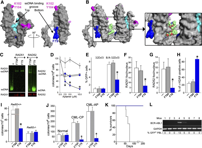
Figure 3.
F79 aptamer disrupts RAD52-ssDNA binding and inhibits HRR to elevate the number of lethal DSBs and eradicate CML. (A) Surface views of the RAD52(1-212) protomer; the top and the bottom of the ssDNA binding groove are marked with arrows. Amino acids forming the ssDNA binding groove (DNA I) are colored in light/dark blue and these binding to dsDNA (DNA II) are in magenta. The location of amino acids V71-G83, which form the F79 aptamer, is highlighted in dark blue. All structures were created using the PyMOL program. (B) The F79 aptamer surface (light green) is shown between 2 RAD52 monomers to illustrate the size of the aptamer and demonstrate that it is a better fit to the binding groove than the other RAD52 monomer. The zoomed box focuses on the area that the aptamer occupies between the 2 RAD52 monomers. (C) F79 and F79A aptamers were added to the mixture of IRDye800-ssDNA and GST-RAD52 protein (right) or IRDye800-ssDNA and GST-RAD51 protein (left); the presence of ssDNA-RAD52 and ssDNA-RAD51 complexes were detected by agarose fluorescent gel shift assay (upper) combined with western blotting (lower). (D) Number of colonies from normal and CML-CP Lin−CD34+ cells (circles and triangles, respectively) incubated with the indicated concentrations of F79A (gray) and F79 (blue) aptamer; *P < .001 in comparison with F79A. (E-H) Cells were untreated (white bars) or treated with F79A (gray bars) and F79 (blue bars) aptamer. (E) Percentage of GFP+ cells representing HRR activity in 32Dcl3 and BCR-ABL1 (B/A)-32Dcl3 cells; *P = .01. (F) Number of RAD51 foci/nucleus in Lin−CD34+ CML-CP; *P < .001. (G) Number of RAD52 foci/nucleus in Lin−CD34+ CML-CP; *P < .001. (H) Percentage of Lin−CD34+ CML-CP cells containing more than 20 γ-H2AX foci/nucleus; *P < .001. (I) Number of colonies from BCR-ABL1 Rad52+/+ and Rad52−/− cells incubated with aptamers; *P < .001 in comparison with untreated and F79A group. (J) Clonogenic activity of Lin−CD34+ cells from 3 healthy donors and 3 CML-CP and 3 CML-AP patients incubated with aptamers; *P < .001 in comparison with untreated counterparts. (K) Survival of nonobese diabetic/SCID mice bearing BCR-ABL1 Rad52+/+ leukemia and treated with F79 and F79 aptamers (8 mice/group). (L) RT-PCR detection of BCR-ABL1 mRNA in bone marrow mononuclear cells from mice injected with BCR-ABL1 Rad52+/+ leukemia cells and subsequently treated with F79 aptamer, which survived more than 200 days. GAPDH served as positive control. %GFP+ PBL, percentage of GFP+ BCR-ABL1 leukemia cells detected in peripheral blood mononuclear cells.

