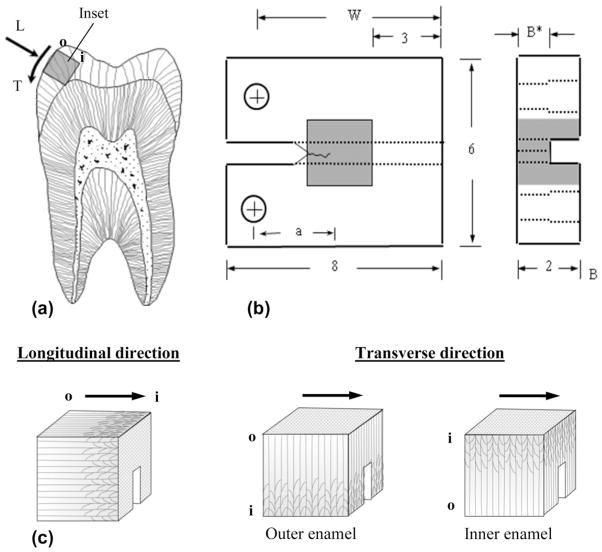
Fig. 1.
Specimen preparation, geometry and testing orientations. (a) Buccal–lingual section of molar with potential inset of occlusal enamel highlighted. Note the longitudinal (L) and transverse (T) directions of crack growth. The letters “o” and “i” refer to the outer and inner enamel, respectively. (b) Geometry of the CT specimen. The molded enamel inset is approximately 2 × 2 × 2mm3 and embodied within a dental resin composite. All dimensions are in millimeters. (c) Relative placement of the back channel in enamel inset for the longitudinal and transverse orientations directions. The arrows illustrate the path of crack growth. For the transverse direction, samples are shown for crack growth within the outer and inner enamel as indicated.

